Sunday, October 6, 2013

Sensation & Perception

KEY TERMS:

Sensation - detecting stimuli from the body or environment

Perception - organizing sensations into meaningful patterns

Stimulus - Form of energy that can affect sense organs

Psychophysics  - study the relationship between stimuli & our psychological response to them

Sensory receptors - cells designed to serve very specific functions such as to detect stimuli & convert energy into neural impulses

Thresholds - a minimum amount of any given sensation that has to be present for us to notice it

Absolute threshold -This is the minimum amount of a stimulus that is necessary for us to notice it 50% of the time

Sensory adaptation - If a stimulus is unchanging, we become desensitized to it. Keeps us focused on changes, not constants

Just noticeable difference (JND) - smallest difference in amount of stimulation that a specific sense can detect

Weber’s law - states that the size of the JND is a constant proportion of the initial stimulus

VISION 

Hue determines color
Intensity determines brightness
Transduction - the process where the eye converts electromagnetic energy (light) into nerve impulses

The Eye 


Parts: 
Cornea - light is initially focused by this transparent covering over the eye
Pupil - Light enters the eye through this opening
Iris - Muscle connected to the pupil that changes its size to let in more or less light
TRIVIA: Everyone has a unique iris (thus it is a new security technique being employed by some organizations)
Lens - This flexible disk under the cornea focuses light onto the back of the eye. It has capacity of
Accommodation - Flexibility of the lens allows eye muscles to adjust light from objects at various distances away
Retina - Light reflected from the lens is received by this sheet of tissue at the back of the eye; it contains the receptors that convert light to nerve impulses
Cones - retinal cells that respond to particular wavelengths of light, allowing us to see color and are located mostly on the fovea, which gives us the sharpest resolution of visual stimuli
          Theories on Seeing Colors
         A. Trichromatic theory - our eyes have three types of sensors (Red, blue & green receptors)
         B. Opponent-process theory - receptors respond to pairs of colors (White-black / red-green / yellow-blue)
Rods - retinal cells that are very sensitive to light but only register shades of gray (i.e., no color); are located everywhere in the retina except in the fovea (we see best at night without light in the periphery of our vision) and allow us to see at night without strong light (this is why we see less color at night)
Optic Nerve - this is where the converted impulse from light is directed from the receptor cells in the retina; it is a large bundle of nerve fibers that carry impulses from the retina to the brain and sits on the retina but contains no cones or rods, so this is where you experience a ‘blind spot’
(TRIVIA: We aren’t aware that we have a blind spot because our brain completes patterns that fall across our blind spot and because our eyes are constantly moving (‘filling’ it in) as discussed in Gestalt Principles of Perception.)

Processing of Visual Information
The retina processes electrical impulses. It encodes and analyzes sensory information (at the most basic level) then in the optic nerve, the neurons pick up the messages from retina, transmit to the thalamus, then on to the visual cortex, then on to more specified areas.

Gestalt Principles of Vision
Figure-ground - we recognize figures (objects) by distinguishing them from the background
Proximity - Marks that are near one another tend to be grouped together
Closure - we tend to fill in gaps in a figure
Similarity - Marks that look alike tend to be grouped together
Continuity - Marks that tend to fall along a smooth curve or a straight line tend to be grouped together

Depth Perception - the ability to perceive a 3-dimensional object when our eyes only project a 2-dimensional image on our retinas
Different cues to perceive depth:
Binocular disparity - since we use both our eyes to focus on an image, the angles used by each eye to put the image on the fovea of our retina is used by the brain to perceive distance
Monocular cues - Our brain also uses information from the stimulus that does not involve our use of both eyes
Motion - specifies distance of an object based on its movement
Motion parallax - objects that are closer to us move farther across our field of view than do objects that are in the distance
Texture gradient - progressive changes in surface texture that signal distance
Linear perspective - parallel objects seem to get closer together as they get farther away

Perceptual Constancy - The image of an object on your retina can very in size, shape, and brightness but we still continue to perceive the object as stable in size, shape and brightness
Size constancy - The tendency to view an object as constant in size despite changes in the size of its image on the retina (as we move)
Shape constancy - The tendency to see an object as retaining its form despite changes in orientation
Color constancy - The tendency to view an object as retaining its color despite changes of brightness in the environment

AUDITION

Pitch - Frequency of air waves
Loudness/volume - Amplitude of air waves

The Ear

http://www.ndt-ed.org/EducationResources/HighSchool/Sound/Graphics/Ear.gif
Outer ear: Pinna
& external auditory canal
Middle ear: Eardrum (tympanic membrane), hammer, anvil & stirrup
Inner ear: Oval window, cochlea (basilar membrane inside)

The Hearing Process
Air waves move the tympanic membrane (eardrum), which moves the hammer, anvil and stirrup (these all amplify the air wave and pass it on)to the basilar membrane in the cochlea. Here, different frequencies are transduced via hair cells (i.e., the receptors of the ear) into nerve impulses that are sent to the auditory cortex of the brain.

Theories on Hearing
There are two theories on transduction in basilar membrane (i.e., how we perceive sound):
Frequency Theory states that neural impulses are stimulated more with higher frequencies of air waves
More plausible for small frequencies, rather than high frequencies because we can hear freqs higher than the maximum rate of neural firing (1,000 neurons a second)
Place Theory states that different frequencies of air waves activate different places along the basilar membrane

TACTILE - SENSE OF TOUCH

  • Skin is the body’s largest sensory organ. Millions of skin receptors mix and match to produce specific perception. 
  • Four basic types of sensations are: Pressure, warmth, cold, and pain. Skin senses pain and warns us of impending danger. 
  • Our brain releases endorphins which are neurotransmitters that have a pain-killing effect.
  • Gate-control theory explains that pain impulses can be inhibited by closing of neural gates in the spinal cord. 


Kinesthetic sense 


  •  provides info about position of joints, muscles, limbs; gives us control over body movements


Vestibular sense 


  •  provides info about body’s orientation relative to gravity and head’s position in space; helps us maintain balance
  • relies on semicircular canals in the inner ear

Olfaction 


  • sense of smell through detecting molecules in the air  
  • Olfactory receptors are built so that only molecules with particular shapes will fit in particular receptors
  • Receptors send neural signals to the brain, passing the thalamus (memory) and the limbic system (emotions) along the way this is why odors often trigger emotional memories 

Gustation 


  • sense of taste through detecting molecules of substances that have dissolved in saliva
  • the sense of taste combines with the sense of smell to produce perception of flavor of food
  • Research suggests that neural impulses for both senses converge to some degree in brain area associated with the perception of flavor
  • When the sense of smell is blocked, we have a harder time detecting most flavors

Taste buds - clusters of hair-like receptor cells; within each bud is a cluster of 50 to 150 receptor cells
TRIVIA: We have about 10,000 taste receptor cells (most on tongue)
Four types of taste: sweet, sour, salty & bitter


References:

Myers, D. (2005) General Psychology

Biological Basis of Behavior Part 2: (Endocrine System)

Endocrine System

 - collection of glands that produce hormones that regulate body's growth, metabolism, and sexual development and function

Functions:
a) Secrete affective hormones directly into the bloodstream;
b) Regulate hormone secretion to normal levels needed by the body and;
c) Maintain interaction with the central nervous system to promote normal body functioning.

Key parts:
a) Pituitary Gland – primarily dubbed as the “Master Gland”, it controls the activities of the other endocrine glands. It secretes growth hormones (crucial for controlling body growth) and an antidiuretic hormone (controls water excretion through the kidney)
b) Thyroid Gland – it is located at the base of the throat and is responsible for the secretion of hormones that control basal metabolism.
c) Adrenal Glands – These are located on top of each kidney. The glands’ inner layer secretes adrenaline or epinephrine, which gives the body energy in times of stress. The outer layer is responsible for the production of cortical hormones (stress hormones).
d) Islet of Langerhans  – This gland is located in the pancreas which is responsible for the secretion of insulin that prevents the accumulation of sugar in the blood.
e) Gonads – The testes for males and the ovaries for females consecutively make up the gonads or sex glands. The former secretes testosterone and the latter secretes estrogen and progesterone.

Associated Illnesses/Disorders
- caused by either the oversecretion or undersecretion of the hormones of the glands mentioned above.
a) Oversecretion –
   Giantism (growth hormone)
   Hyperthyroidism (metabolic hormones)
   Extreme body weakness (Insulin)
b) Undersecretion –
   Dwarfism (growth hormone)
   Hypothyroidism (metabolic hormones)
   Diabetes (Insulin)

http://www.inspirationgreen.com/assets/images/Issues/2013/endocrine-glands-and-their-hormones.jpg

The Biological Basis of Behavior: Part 1 (Nervous System)


CONTENTS:
       I. The Nervous System
       II. Cells in the Nervous System
 III. Neurotransmitters

DISCUSSION: 
I. The Nervous System
      The nervous system is the body’s electrochemical communication circuitry. The field that studies the nervous system is called neuroscience, and the people who study it are called neuroscientist.

      Characteristics of Nervous System
      a. Complexity - due to the orchestration of the billion of cells in the brain and nervous system, the individual can do complex or different kinds of activities.
      b. Integration - the ability of the brain to pull information together
      c. Adaptability - although the composition of the brain and the nervous system have hereditary       foundation, both have the ability to constantly adapt to the changes in the body and the environment.   
      d. Plasticity - denotes the brain’s special capacity modification and change. 
      e. Electrochemical transmission - the brain being the information processing system, powered by electrical impulses and chemical messages allows the individual to perceive and respond stimuli.

     Organization of the Nervous System
     The nervous system is organized into two main parts:
     1. The central nervous system (CNS), encased in bone, consists of the brain and spinal cord. The CNS is the nervous system’s central executive.
     2. The peripheral nervous system extends throughout the body and relays information to and from the brain.

http://classes.psy.ohio-state.edu/100/upload/farmer/images/nervoussystemorganizationchart.jpg

      I.A.  The Central Nervous System
     The CNS performs different functions through different networks of neurons. Clusters of neurons are called nuclei and pathways that connect the networks are bundles of axons called fiber tracts.
     I.A.1. The Brain 
     1. The Hindbrain - is found just above the spinal cord and is composed of the following structures:
           a. The medulla controls vital life functions (e.g., blood pressure, heart rate, and breathing).
           b. The reticular formation is a web of neurons is involved in arousal and attention.
           c. The cerebellum coordinates fine motor movements, stores a memory code for well-rehearsed   
               behaviors, and participates in cognitive tasks such as reading.
    2. The Midbrain - relays information from the eyes, ears, and skin and controls certain types of automatic behaviors. The midbrain and its connections to the forebrain permit the smooth initiation of movement. The midbrain is connected to the brainstem which is the posterior part of the brain, adjoining and structurally continuous with the spinal cord. Reticular formation is a region in the brainstem that is involved in multiple tasks such as regulating the sleep-wake cycle and filtering incoming stimuli to discriminate irrelevant background stimuli.
    3. The Forebrain - the largest part of the brain regulates many complex aspects of behavior and mental phenomena. Interior structures include the following:
        a. thalamus- processes inputs from sense organs (except for smell) and then relays sensory information to appropriate “higher” forebrain areas. It is the primary sensory relay into the rest of the brain. The brain’s “clock” that sets biological rhythms for the body.
      b. hypothalamus- is a portion of the brain that contains a number of small nuclei with a variety of functions. Its functions are:  (1) link the nervous system to the endocrine system via the pituitary gland (hypophysis) and; (2) control body temperature, hunger, important aspects of parenting and attachment behaviors, thirst, fatigue, sleep, and circadian cycles.
      c. The limbic system includes the amygdala and the hippocampus. The amygdala is involved in memory and emotion. It links different kinds of sensory information together in memory. The amygdala also plays a role in fear and other emotions, linking emotions to sensations. The hippocampus is critical to the ability to form new memories.
      d. The cerebral cortex, is a thin sheet of neurons comprising the forebrain’s outer surface. It folds in itself, giving the brain a wrinkled appearance. The cerebral cortex is divided down the middle, creating two halves called the left and right cerebral hemispheres. The corpus callosum connects the two halves. The folds of cortex produce gyri (ridges) and sulci,or fissures (valleys or wrinkles), on the brain’s outer surface. Several deep sulci make convenient markers for dividing the cortex of each hemisphere into four anatomical areas: the frontal, parietal, occipital and temporal lobes.
         d. 1. The Frontal Lobe of the brain is located deep to the Frontal Bone of the skull. It plays an integral role in the following functions/actions: Memory formation, Emotions, decision making/reasoning and personality.
           d. 2. The Parietal Lobe of the brain is located deep to the Parietal Bone of the skull. It plays a major role in the following functions/actions: Senses and integrates sensation(s), Spatial awareness and perception (Proprioception - Awareness of body/ body parts in space and in relation to each other).
        d. 3. Occipital Lobe involves two major parts: (1) Primary Visual Cortex – the primary area of the brain responsible for sight -recognition of size, color, light, motion, dimensions, etc; and (2) Visual Association Area – interprets information acquired through the primary visual cortex.
           d. 4. The Temporal Lobes are located on the sides of the brain, deep to the temporal bones of the skull. They play an integral role in the following functions: Hearing, Organization/Comprehension of language, Information Retrieval  (Memory and Memory Formation).
      I.A.2.The Spinal Cord
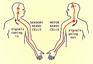
      It receives and sends signals to and from the brain. There are 31 pairs of spinal nerves which run through the spinal cord. These nerves are called “mixed” nerves because each nerve contains a sensory and a motor axon. The spinal cord can also be a minor coordinating centre for some simple reflexes like the withdrawal reflex.
http://www.siumed.edu/~dking2/ssb/brainday/inout.jpg

Meanwhile, reflexes are simple, involuntary behaviors controlled by spinal cord neurons, without requiring instructions from the brain. These are controlled by a feedback system. Information about the consequences of an action goes back to the source of the action for further adjustment, if necessary.

     I.B. The Peripheral Nervous System
     It has two subsystems:
      I. B. 1. The Somatic Nervous System - carries signals between the senses and CNS and between the CNS and skeletal muscles. Sensory neurons bring information to the brain, and motor neurons send information from the brain to the muscles.
      I. B. 2. The Autonomic Nervous System - carries messages between the CNS and the heart, lungs, and other organs and glands. The ANS has two divisions that may act on the same body areas, with their relative “balance” regulating the state of the targeted organs:
         I.B.2.a. The sympathetic system directs the body to spend energy (e.g., increased heart rate, faster breathing, sweating, sometimes called the fight-or-flight” response) to react to stress.
         I.B.2.b. The parasympathetic system directs the body’s functions to conserve energy (e.g., slower heart rate, increased digestive activity). Parasympathetic activity helps “calm” a person after increased sympathetic arousal.

II. Cells in the Nervous System
There are two main cell types in the nervous system. Neurons (also known as nerve cells) are specialized to respond rapidly to signals and send signals of their own while glial cells provide energy, help restore damage, and respond to signals from neurons. These cells have some features in common. They both have an outer membrane that selectively allows only some substances to pass in and out. The only notable differences between neurons and glial cells are neurons' possession of axons and dendrites, and capacity to generate action potentials.
  
Moreover, neurons have: cell body (also known as soma) which contains the nucleus; mitochondria turn oxygen and glucose into energy; axon which is a cell fiber that carries signals away from the cell body and a dendrite which is a cell fiber that receives signals from other neurons and carries information toward the neuron’s cell body. Most neurons have one axon but have many dendrites. Some axons are wrapped in a myelin sheath formed from the plasma membranes of specialized glial cells known as Schwann cells which serve as supportive, nutritive, and service facilities for neurons. The gap between Schwann cells is known as the node of Ranvier, and serves as points along the neuron for generating a signal.
http://brainu.org/files/tn_about_neurons.jpg


As mentioned, neurons have special features that permit effective signal communication and they have the capacity to generate action potentials. Action potentials are electrochemical pulses that shoot down the neuron’s axon. They are “all-or-none” which means that a neuron either fires an action potential at full strength or does not fire at all. After an action potential, there is a brief recovery time called a refractory period, during which a neuron cannot fire another action potential. The speed of an action potential depends on the thickness of the axon and on the presence of myelin sheath, a white, fatty substance that speeds up action potentials. At the axon endings, the action potential causes bag-like vesicles to release stored chemicals called neurotransmitters into a space between the two neurons. This space is called a synapse, a connection that is a narrow gap separating the axon of one neuron from the dendrites of another. It is the means by which two neurons communicate. Released neurotransmitters “float” across the synapse to “bind” with receptors and proteins on a dendrite of a receiving neuron. The interaction between neurotransmitters and receptors is very specific, like a lock and key.  This interaction creates a signal called a postsynaptic potential (PSP) that might make action potentials in the receiving, or postsynaptic, neuron either more or less likely. A number of PSPs sum together at the junction of the cell body and the axon. Whether or not an action potential “fires” depends on the kind of signals that are most numerous.

Neurons have three kinds (as taken from wikipedia.com):
1. Sensory neurons are neurons responsible for converting various external stimuli that come from the environment into corresponding internal stimuli. They are activated by sensory input, and send projections to other elements of the nervous system, ultimately conveying sensory information to the brain or spinal cord. Unlike neurons of the central nervous system, whose inputs come from other neurons, sensory neurons are activated by physical modalities such as visible light, sound, heat, physical contact, etc., or by chemical signals for example in the case of smell or taste.
2. Motor neuron (or motoneuron) classically applies to neurons located in the central nervous system (CNS) that project their axons outside the CNS to directly or indirectly control muscles. Motor neurons are efferent nerves also called effector neurons that carry signals from the spinal cord to the muscles to produce (effect) movement.
3. An interneuron (also called relay neuron, association neuron, connector neuron or local circuit neuron) is a neuron that forms a connection between other neurons. Interneurons are neither motor nor sensory. The term is also applied to brain and spinal cord neurons whose axons connect only with nearby neurons, to distinguish them from "projection" neurons, whose axons (projection fibers) project to more distant regions of the brain or spinal cord.

http://images.tutorvista.com/content/nervous-coordination/types-of-neurons.jpeg
      III. Neurotransmitters
    There are about 100 neurotransmitters that have been identified (which means some are still undiscovered). A group of neurons that communicate using the same neurotransmitter is called a neurotransmitter system.  Neurotransmitters are chemical messengers that traverse the synaptic gaps between neurons. When released, these travel across the synapse and bind to receptor sites on the receiving neuron, thereby influencing whether it will generate a neural impulse. Below are some of its types:
a) Acetylcholine is used by sets of neurons involved in controlling movement of the body, in making memories, and in slowing the heartbeat and activating the digestive system. Alzheimer’s disease may result from disruptions of this system.
b)  Norepinephrine affects arousal, wakefulness, learning, and mood. Disruptions of this system have been linked to depression.
c) Serotonin affects sleep, mood, aggression, and impulsive behaviors. Serotonin levels can be affected by what is eaten.
(1) Malfunctions in serotonin systems can result in mood and appetite problems seen in some types of obesity, premenstrual tension, and depression.
 (2) Antidepressant medications such as Prozac, Zoloft, and Paxil are thought to act on serotonin systems to relieve some of the symptoms of depression.
d) Dopamine is used by sets of neurons involved in controlling movement, and damage to these systems contributes to shakiness experienced by people with Parkinson’s disease. Other dopamine systems are involved in the experiencing of reward, or pleasure, which is vital in shaping and motivating behavior. Certain other dopamine systems are suspected to be responsible for the perceptual, emotional, and thought disturbances associated with schizophrenia.
e) GABA (gamma-amino butyric acid) is the main inhibitory neurotransmitter in the brain—it slows down the brain’s neural activity.
(1) Some drugs amplify the inhibitory action of GABA. One example is alcohol, which results in impairments of thinking, judgment, and motor skills. Drugs that interfere with GABA’s inhibitory effects produce intense repetitive electrical discharges, known as seizures
(2) Impaired GABA systems are thought to contribute to severe anxiety, Huntington’s disease, and epilepsy.
f. Endorphins - natural opiates that mainly stimulates the firing of neurons. It shields the body from pain and elevates feelings of pleasure.


References: 


Carlson, N. R. (1997). Psychology: the science of behavior. Allyn & Bacon
Gazzaniga, M. S., Ivry, R. B., and Mangun, G. R. (1998). Cognitive neuroscience: the biology of the mind. New York, NY: W. W. Norton & Company, Inc.
Goldstein, E. B. (1999). Sensation and perception (5th ed.). Pacific Grove, CA:Brooks/Cole Pub.
Kolb, B. and Wishaw, I.Q. (1996). Fundamentals of human neuropsychology (4th ed.). New York, NY: Freeman.
Rosenzweig, M. R., Leiman, A.L., and Breedlove, S. M. (1999). Biological psychology: An introduction to behavioral, cognitive, and clinical neuroscience (2nd ed.). Sunderland, MA: Sinauer Associates, Inc.
http://serendip.brynmawr.edu/- Articles and links on the brain and behaviour.
http://www.neuroguide.com/ - Links to journals, images, and  resources.
http://anatomy.umas.edu/HTMLpages/anatomyhtml/neuro_atl as.html – Complete pictorial atlas of the brain.
http://www.cc.emory.edu/ANATOMY/AnatomyManual/nervous_system.html – Illustrated tutorial of the nervous system
http://en.wikipedia.org/wiki/Neuroglia 
http://www2.estrellamountain.edu/faculty/farabee/biobk/biobooknerv.html

Sunday, June 9, 2013

Course Outline

Week 1            
I. Introduction to Psychology                                                                              
A.       Its Definition and Meaning
B.      Brief History and Notable Psychologists
C.      Development of Psychology in the Philippines
D.      Goals and Significance of the Studying Psychology
E.       Branches and Related Fields of Psychology

Week 2
II. Principles of Growth and Development                                                                      
        A.  Reproduction Process
        B.  Genetics and Heredity
1. Ten Stages of Human Development
2. Aspects of Human Development
      a. Motor
      b. Social
      c. Cognitive
      d. Moral
      e. Psychosexual
      f. Psychosocial

Week 3
FIRST LONG EXAMINATION                                                                                   

Week  4 & 5
III. Biological Bases of Behavior                                                                                   
A. The Nervous System
1. Central Nervous System
2. Peripheral Nervous System
B.  Endocrine Glands
  
 Week 6 & 7
IV. Sensation & Perception                                                                                                    
           A. Sensation                                                                                                        
1.    The Different Sensory System
                   a. Visual
                   b. Auditory
                   c. Olfactory
                   d. Gustatory
                   e. Kinesthetic
                   f. Vestibular
    2. Perception                                                                                                         
              a. Perceptual Organization
              b. Depth Perception
              c. Perceptual Constancy
              d. Illusions
              e. Major Influence on Perceptual Processes
              f. Barriers to Accurate Perception of Others

Week 8
MIDTERM EXAMINATION                                                                                          

Week 9 & 10 
V. Learning and Memory                                                                                                    
          A. Learning                                                                                                            
            1. Definition
            2. Theories in Learning
                 a. Classical Conditioning
                 b. Operant Conditioning
                 c. Connectionism
           B. Memory                                                                                                        
            1. Definition
            2. Basic Types of Memory
            3. Information Processing Model
            4. Forgetting
                    a. Theories of Forgetting

Week 11 
VI. Intelligence and Measurement                                                                  
            A. Levels of Intelligence
            B. Multiple Intelligence

Week 12
SECOND LONG EXAMINATION                                                                                

Week 13 & 14
VII. Motivation and Emotion                                                                                               
A. Motivation                                                                                                       
1. Theories of Motivation
            2. Classification of Motivation
          B. Emotion                                                                                                          
                  1. The Nature and Physiology of Emotion
                  2. Theories of Emotion
                  3.  Classifications of Emotion
                  4.  Emotional Quotient (EQ)

Week 15 & 16
VIII. Individual Differences                                                                                                              
 A. Personality                                                                           
               1. Nature of Personality
               2. Theories of Personality
                        a.  Psychoanalytic
                        b.  Personology
                        c.  Factor Analysis
                        d.  Perceived Reality
                        e.  Person Centered 
                        f.  Somatotypes
  B.  Abnormal Behavior                                                                               
                   1. Definition
                   2. Classification and Categories
                   3. Psychological Assessment

Week 18 
FINAL EXAMINATION   

XXX

(adapted from the prescribed General Psychology Syllabus, CvSU)